Barium Enema
What Is Barium Enema And What To Expect If You Are Having One
A barium enema is a type of test done by introducing a dye called barium into the large intestine or colon through the rectum and then an X-ray picture is taken of the lower digestive system to reveal any abnormality which would be enhanced by the dye. See why it is done, how it is done, what to expect and preparations you must make before going for the test.
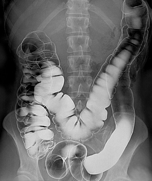
 Barium enema film. The white dye filling the outline of the large intestine is the barium.
A barium enema is usually done to take a look at an impression or filling defect that any abnormality in our large intestine may leave when filled up with barium.
The large intestine or colon is:
- That part of our gut that absorbs the water that is left in food you have eaten that is still left after passing through the upper digestive system.
- The undigested food that is left is excreted through the rectum as a waste product.
- The large intestine is a muscular tube, and it is only one quarter as long as the small intestine, but it has a larger diameter of about 7.5 centimeters wide at the cecum and it narrows to approximately 2.5 centimeters at the sigmoid colon, before it becomes the rectum, a structure that is about 10 centimeters long in adults.
- The large intestine has cells which line the interior and these cells secrete and absorb fluid. There are also immune cells in the intestine, as well as normal bacteria.
- These bacteria have a useful function to play, such as making vitamins like vitamin K, and if antibiotics kill them off, a very bad type of bacteria might take over and cause diarrhea and even rupture of the colon.
- The large intestine also neutralizes acids that form in the earlier parts of the digestive process, and the walls of the intestine act as a barrier to protect against the invasion of bacteria into the bloodstream.
- Finally, some antibodies, substances that our body produces to fight harmful substances and disease, are made in the large intestine.
Why A Barium Enema?

Barium enema is still a very useful test for some conditions today, despite the availability of CT scan and other tests. Since it comes with the advantage of requiring lesser exposure to radiation compared to doing a CT scan and being less invasive too, compared to a colonoscopy it is still very much commonly ordered by doctors across the world.
It is also less expensive than most other test.
Your doctor might order a barium enema to:
- Find evidence of inflammation and irritation of the wall of the intestine that occurs in diseases like Crohn’s disease or ulcerative colitis. If you have one of these diseases, your doctor may monitor your progress by having barium enema tests done at regular intervals.
- Sometimes there are problems with the structure of the large intestine. These problems may include little outpunching pockets that are called diverticula, which can fill with food waste and become inflamed.
- Other problems with the structure of the large intestine may include narrowed areas that are called strictures. They can be seen on a barium enema image study.
- There are a few conditions where the intestine may twist or where one portion may slide up into the lumen, or middle, of another section, and this is called intussusception. Sometimes a barium enema will cure this problem just by doing the test.
- If you have had abdominal pain that can’t be explained, blood in your stool, changes in your bowel movements, unusual weight loss, or anemia, your doctor will want to do a barium enema to see if you have any unusual growths like tumors that may be malignant, or polyps which may be pre-malignant.
How A Barium Enema Is Done - What To Expect
A barium enema uses a contrast material with barium in it, and the barium blocks the X-rays, which make the image clearer.
- The barium is given by putting the contrast material through a tube, which is inserted into the anus, at the very end of the rectum.
- The contrast material fills the colons, and makes an outlined image of the colon, which can make large abnormalities show up well on the X-ray of the colon. This is called a single contrast barium enema.
- Sometimes, the colon is filled with barium, which is then drained out. There is then a thin coating of barium on the wall of the intestine.
- The second part of this procedure involves next filling the intestine with air. This is called a double contrast barium enema. This type of study allows the doctor to see more details of the inner surface of the colon, particularly in the case of diverticula, narrowing strictures, or inflammation.
The single contrast barium enema is often done either for a specific medical purpose or because some people may be too old or too ill to be comfortable during the longer process of the double contrast study.
However, if the results of the single contrast study are not able to determine if there is an abnormality, the patient may have to have a double contrast study after all.
When you arrive for your test:
- You will be taken to an X-ray suite, and your will be given a gown to change into. The doctor will put the barium through the anus with a tube.
- You will first lie flat while an X-ray is taken, and when you turn onto your side the enema will be inserted gently.
- If you have cramping, you may receive some medication to help with the crampy pain.
- You may have to turn one way and back again as your doctor watches the barium flow through your intestine through his X-ray fluoroscope monitor.
A single contrast barium enema may take 30 to 45 minutes, and the double contrast type of scan may take as long as an hour, but the barium will only be inside for ten to fifteen minutes of that time.
When the barium enema examination is over, the enema tube is removed, and you can use a bedpan or toilet to expel the barium. A few additional X-rays may be taken without the barium at this point.
How Do I Prepare for a Barium Enema?
Before you have a barium enema, you must answer some very important questions for your doctor. These include telling your doctor about any of the following:
- If you are allergic to latex
- If you are allergic to barium
- If you are pregnant or if you may have the possibility of being pregnant
- If you have had another barium test in the upper portion of the digestive tract recently
- If you have had a colonoscopy or a sigmoidoscopy recently
Cleansing the bowel for a barium enema.
When you have a barium enema, you have to prepare by completely cleaning out your large intestine, because any stool will make the test less accurate.
- You will only be able to drink clear liquids, clear soups, or water for one to three days before your study.
- Beginning 24 hours before the test, your doctor will probably tell you to be drinking large amounts of clear liquids, which are not carbonated.
- You will have to take some laxatives prescribed by your doctor to empty out your intestines.
- Sometimes, your doctor will ask you to give your self an enema with tap water to completely clear out the intestine.
You may have to repeat this twice until the water that you pass is clear of particles. Your doctor may give you a suppository instead, or a Fleet enema.
What Happens After My Test?
After your barium enema test, your doctor will remind you to drink a lot of water to replace all of the fluid you lost when you were cleaning your intestines.
He may also tell you to use a laxative to get all of the barium out of the intestine. You may have some pain around your anal region from the preparation and from the barium enema itself. You can sit in a warm bath to relieve any discomfort, and a salve like Preparation H may be helpful.
The doctor who ordered the test for you may not be the radiologist or the gastroenterologist who does the test, so your doctor will get the results and will let you know as soon as he has the results in hand.
References:
- Fischbach FT, Dunning MB III, eds. (2009). Manual of Laboratory and Diagnostic Tests, 8th ed. Philadelphia: Lippincott Williams and Wilkins.
- Pagana KD, Pagana TJ (2010). Mosby’s Manual of Diagnostic and Laboratory Tests, 4th ed. St. Louis: Mosby Elsevier.


