Pelvic Ultrasound Scan
Understanding Pelvic Ultrasound Scan - Uses, Preparations And Results
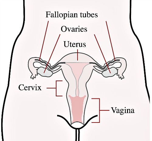
What Is A Pelvic Scan?
 Pelvic ultrasound is the use of a machine that gives off sound waves to generate a picture of the structures inside the pelvis of a man or woman. It is the ultrasound scan of the lower abdomen and reproductive area.
The pelvis is the part of the body, which contains your genital organs, the bladder and the reproductive organs in women, and the prostate gland and bladder in men. Usually, pelvic ultrasounds are only done in women.
An ultrasound is a diagnostic machine that uses sound waves that are at a frequency too high for humans to hear. These sound waves are projected into the body from a probe that is called a transducer. Because it takes the sound waves longer or shorter lengths of time to travel through different body tissues, depending upon how much water the tissue has, the reflection of the sound waves can be used to create two dimensional images of organs and spaces within the body, from the outside.
There is no radiation from ultrasound, so it is very safe. It is also very portable and inexpensive.
There are two types of pelvic ultrasound examinations.
One is done with the probe on top of the abdomen. There is some jelly placed on the probe to conduct the sound waves. The other type of examination is done using a smooth probe that fits in the vagina. This type of examination can often get better pictures of the organs in the pelvis compared to the abdominal ultrasound.
The type of ultrasound examination ordered by your
doctor depends upon the purpose of the study he wants, the organs he wants to
see clearly, and the reason for the test.
Why Would My Doctor Be Requesting A Pelvic Ultrasound Scan?
A primary use of the pelvic ultrasound examination is during pregnancy, when it is used to determine the size of the fetus and to compare that size to the estimated ages of gestation, which is determined by the date of your last menstrual cycle. The technician can determine the size of the fetus by taking measurements with the mouse on the ultrasound computer.
As your pregnancy advances, you will have additional ultrasound studies in order to be certain that the fetus is growing and that the fetal heartbeat is present. Your doctor should be able to see the fetal heartbeat about 6 weeks after conception.
The doctor will also look for any fetal abnormalities, which may be things like neural tube defects, which occur when the vertebrae, or small bones of the back, fail to develop properly. If the vertebrae do not develop fully, the infant will have no way to protect the spinal cord.
The doctor will also want to check to be certain that there is enough amniotic fluid, which is the fluid in the uterus that serves as protection for the developing baby.
During your pregnancy, you may decide to have a test called amniocentesis, during which the doctor inserts a large needle into the amniotic sac, which contains the fetus and the amniotic fluid. Your doctor will take a sample of the amniotic fluid and may take a sample of the placenta, through which the fetus receives nutrients and oxygenated blood from the mother. These tests are done to see if there are any problems with the genetic development of the fetus, and the tests are called amniocentesis and chorionic villae sampling. The ultrasound is necessary to guide the doctor as he inserts the needle into the amniotic sac.
If you are a woman of childbearing age, and you are having abdominal pain, the doctor in the clinic or emergency department will perform a pelvic ultrasound examination if you have a positive pregnancy test and have not already had a pelvic ultrasound performed.
Your doctor will have to be certain that your pregnancy is developing inside the uterus. Abdominal pain in the case of a positive pregnancy test can potentially be a pregnancy that is developing outside of the uterus. This type of pregnancy is called an ectopic pregnancy, or tubal pregnancy, and if you are having abdominal pain with an ectopic pregnancy, it could indicate that the ectopic pregnancy is causing irritation and may rupture. If that happens, you could require surgery.
If you are not pregnant, there are still many other reasons that your doctor may order a pelvic ultrasound. Certain symptoms may cause your doctor to want to do a study of the pelvic organs or bladder.
These reasons symptoms include:
Some of the things that your doctor may see during a pelvic ultrasound that will help with making a diagnosis include:
- Cysts on your ovaries, which can cause pain
- Fibroid tumors, which are benign tumors in the wall of the uterus
- Any type of irregular mass, which may represent a cancer
- The volume of urine in left in your bladder after you urinate. This can help your doctor determine a reason for urinary incontinence.
- Your ovaries and fallopian tubes, which may help your doctor make a diagnosis if you are having difficulty getting pregnant and the doctor is looking for causes of infertility.
- Cancerous growths or tumors.
- Signs of pelvic inflammatory disease, which is an infection of the uterus.
- Growth of uterine tissue outside of the uterus, which is a condition known as endometriosis, and can cause pain during the menstrual cycle.
- To look at the thickness of the uterine lining.
- To examine the ovaries and to harvest eggs for fertilization if you are having in-vitro fertilization in order to become pregnant.
- To check for an abscess in the pelvis. An abscess is a collection of pus from an infection, and if you have a pelvic abscess, it will have to be drained in order to rid you of your infection.
What Should I Do To Prepare For A Pelvic Scan?
Usually, you do not require any special preparation done prior to doing a pelvic scan, other than the need to ensure you drink adequate amount of water.
If your doctor or the radiologist wants any further preparation, they would ring you before the test to let you know.
You may have your pelvic ultrasound in the doctor’s office or at the hospital.
The following are basic preparation for an ultrasound scan of the pelvis and what you should expect before the test:
- When you arrive for your appointment, you will be given a gown and you will change your clothes into patient's dress.
- Before the ultrasound, you should tell the doctor if you have had any X-rays with barium or other types of contrast. This can interfere with the ultrasound study.
- Tell your doctor about any allergies to latex, because the ultrasound transducer in the transvaginal ultrasound will have a disposable latex condom over it.
- Before a pelvic ultrasound which is done with an abdominal transducer, you will be asked to fill your bladder with between four and six glasses of water, to help push your intestines out of the pelvis.
- Air in the intestine can interfere with the clarity of the test.
- If you are having a transvaginal ultrasound, you will need to empty your bladder.
- You should not drink anything other than water for four to six hours before the test.
On the day of the test, on arrival to the clinic or hospital, you will be given some time to change into a gown and the technician will take you into an exam room with a table in the room, where you will lie during the test.
The technician will put some gel on your abdomen, which will help the conduction of the sound waves.
If you are having a transvaginal ultrasound, the technician will cover the long, narrow probe with a condom and your doctor will carefully insert the probe into your vagina.
During the study, you will be able to see the internal organs of your pelvis on a small television screen. You may feel some pressure during the examination.
After the pictures have been taken, and
all measurements recorded, you will be given a cloth to wipe the gel from your
abdomen. You can then get dressed and return home, and after a day or so, the
doctor will telephone you with the results.

|
||
| search engine by freefind |
Sign Up For
Our Newsletter
Recent Articles
-
Anxiety stomach Pains caused by possible cancer return
Jul 06, 23 02:54 PM
Diagnosed with Prostate Cancer 16 years ago. Over the last 2 years my PSA results have started to climb, therefore yesterday I had a full body bone scan -
Left upper Quadrant pain for 18 months!
Mar 27, 22 06:55 PM
Hi, About 18 months ago I had to call an ambulance as I had severe pain in my abdomen on the left side. I was discharged and they told me it was an intercostal -
Mesenteric Lymphadenitis - Causes, Symptoms, Diagnosis and Treatment
Sep 19, 20 04:16 PM
 Mesenteric lymphadenitis is a common cause of right-sided abdominal pain in children, that mimics appendicitis. Though more common in children, it can occur in


Disclaimer : The information provided on this website is for educational purposes only. It is not a substitute for seeking a one-to-one professional medical advice and treatment.
ALWAYS consult your doctor or healthcare provider to see if the information provided here applies to your personal situation and for personalized care.
The presence of hyperlinks from this website to external organizations does not constitute an approval, recommendation or endorsement of these organizations. They are provided for convenience only.
ABDOPAIN.com and its affiliates disclaim any and all warranty or liability for the use or lack of use of any and all information on this website. If you have a medical emergency, call your local emergency medical service.